Search Results for: campanula

Campanulas

I cut a stalk of Campanulas (“Canterbury Bells”) from the garden. I laid the bunch gently on my light box for a sequence of high-key captures (below).

Campanula (Canterbury Bells) © Harold Davis

Often, photography of flowers on a light box destroys my models, and this makes me sad. However, in this case the Campanulas were completely intact following photography. So I placed them in a cut crystal vase, and brought them upstairs to enjoy.

It seemed to me that the flowers were pretty special in the vase, but how to capture them? The answer was to use two light boxes, arranged perpendicularly to each other—as in this Light Box Photography in Three Dimensions tutorial video (post-production for a three-dimensional light box image is shown in the follow-up video here).

My Campanulas in a Vase image is shown below following post-production.

Campanulas in a Vase © Harold Davis

Posted in Flowers, Photography

Fantasy in the Key of Campanula

Often when I am through with the primary photography in a light box design, I continue to experiment by adding lines, weaving botanicals, getting closer, and anything else that is fun that I can think of. Fantasy in the Key of Campanula is an example of this kind of improvisation, with the lines of the Campanulas (Bell flower) laid over the original design, then photographed close for a different kind of composition.

Fantasy in the Key of Campanula © Harold Davis

Posted in Flowers, Photography

Campanulas X-Rays

I write this from the train from Frankfurt to Berlin, where Julian K. and I are sitting in a first class car at the all-important table, with the all-important wi-fi connectivity. Since I am connected to the world of the Internet, I can post this!

Campanulas X-Ray © Harold Davis

We are working on processing images from yesterday’s fusion light box and x-ray photography of flowers. These campanulas as arranged took up almost all the space yielded by the size of the sensor of the x-ray machine. I’ve begun to get a bit of a feeling for how the x-ray sensor processes the electromagnetic waves it receives from the emitter, and how to best arrange the subject in the cone shape that is best for this kind of capture. 

Query: Is capture via x-ray photography, or should another term be used? I think “photography” does fit the bill (there is another, more technical vocabulary related to radiology as well). What do you think?

Posted in Photography, X-Ray

Papaver and Campanula

Papaver and Campanula

For a special project, I started with a high-key photo of poppies and bellflowers (Papaver and Campanula). I created the background by scanning and digitally combining two pieces of papyrus and a sheet of rice paper (Ohiri).

Posted in Flowers, Photography

Papaver and Campanula

Papaver and Campanula

Papaver and Campanula, photo by Harold Davis. View this image larger.

This is an image of Papaver Rhoeas and Campanula—or, to use common names, Poppy and Bellflower—both kinds of flowers from my garden.

I place the flowers on a flourescent light box to create a back lit effect and photographed straight down. There’s very little light coming from the front of these flowers. To increase the transparent effect, I sprayed them with water, brushed the water around for even coverage, and pressed the ensemble gently down with a large piece of 1/2 inch plate glass. I removed the glass before taking the photos used to create this composite image.

My technique with this kind of transparent, high-key image is to bracket exposures, all of them biased to the over-exposed side. In this case, I used my Sigma 50mm macro lens. I set the aperture to f/11 and the ISO to 100. Then I made ten exposures, ranging in shutter speeds from 2 seconds to 1/50 of a second.

I started by processing the lightest and brightest exposure (the 2 second one) in Photoshop. Then I gradually layered in detail from the darker versions, using layers and the Paint Brush Tool in Photoshop.

The resulting composite produced the effect I was looking for: the transulence and brightness of a water color with light apparently shining through the partially transparent flowers.

Posted in Flowers, Photography

Topography of Camellia

In the wake of the death of my parents, I created a new garden on the shaded side of our house. This is a mostly ignored narrow strip between our house and the fence that separates us from the sidewalk on the San Ramon side. I anchored this “memorial garden” with three camellia bushes. The central camellia is an espaliered Camellia “Nuncio’s Pearl” (a full blossom is shown at the bottom of this story). 

I surrounded the camellias with anemones and campanulas, all flowering plants that should thrive in this part shade environment. Hopefully, they will eventually carpet the area with blossoms.

We have several hummingbird feeders in this area, and I plan to add a birdbath.

Topography of Camellia © Harold Davis

Topography of Camellia is an abstraction created from one of the first blossoms from this garden. As with a Rorschach, what you see in this image may depend more on you than on me.

Camellia Nuncio’s Pearl © Harold Davis

Posted in Abstractions, Flowers, Photography

Fresh from the Garden

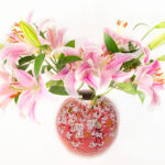
This relatively straightforward yet elegant (if I say so myself) light box composition uses flowers directly from our garden.

Campanulas, Poppies, and a Clematis © Harold Davis

Campanulas, Poppies, and a Clematis © Harold Davis

Posted in Flowers, Photography

Harold Davis—Best of 2018

2018 has been quite a year in art for me. Travel has included the Southwest of France, the Balearic Islands, a pilgrimage on the Camino de Santiago, teaching at Maine Media Workshops, New York City, Heidelberg and Berlin Germany, Paris, Malta, and Sicily. My time at home has been productive as well. 

Below each image, I’ve added links to the relevant blog stories that include my selected images (where I blogged them). Self-selected entries from previous years going back to 2013 can be found here.

Study in Petals on Black © Harold Davis

Blog story: Studies in Petals

Red Anemone © Harold Davis

Blog story: Red Anemone

Devotional Pose © Harold Davis

Blog story: Devotional Pose

Vitruvian Woman © Harold Davis

Blog story: Vitruvian Woman

Egg © Harold Davis

Blog story: Egg

Papaver Nudicaule © Harold Davis

Blog story: Color Field of Flowers

Above the Gran Via © Harold Davis

Blog story: Above the Grand Via

Dandelion in Calvignac A © Harold Davis

Blog story: Dandelion in Calvignac

Bridge Fun © Harold Davis

Blog story: The Art of Being Alone with Oneself

Twisted © Harold Davis

Blog story: Seriously Twisted

Summer Grass © Harold Davis

Blog story: Summer Grass

Poppies and Mallows on White © Harold Davis

Poppies and Mallows on Black (Inversion) © Harold Davis

Blog story: The Art of Photographing Flowers for Transparency

Poppies Dancing © Harold Davis

Poppies Dancing Inversion © Harold Davis

Blog stories: Poppies Dancing and Poppies Dancing on Black

The Passion of the Rose © Harold Davis

Blog story: The Passion of the Rose

Papaver Pod from above © Harold Davis

Blog story: Papaver Poppy Pods Gone to Seed

Poem of the Road © Harold Davis

Lonely Road (Poem of the Road) © Harold Davis

Blog story: Poem of the Road

Sunflower X-Ray Fusion © Harold Davis

X-Ray, Sunflower © Harold Davis

Campanulas X-Ray on White © Harold Davis

Blog story: Revealing the Unseen with X-Ray Photography of Flowers; FAQ: X-Ray Photos of Flowers

Ladyboot Arch © Harold Davis

Blog story: Under the Wide and Starry Sky

Dawn East of the Sierras © Harold Davis

Rising © Harold Davis

Red Pitcher © Harold Davis

Bridge of Light – Color Version © Harold Davis

Bridge of Light © Harold Davis

Blog story: Bridge of Light

Heceta Head Lighthouse © Harold Davis

Heceta Head © Harold Davis

Crepuscular Coast – Black and White © Harold Davis

Crepuscular Coast © Harold Davis

Blog story: Crepuscular Coast

Earthlight © Harold Davis

Blog story: Earthlight

Paris from Montmartre © Harold Davis

Blog story: View of Paris from my room

Paris Paris © Harold Davis

Blog story: Goodbye Paris

Time Machine © Harold Davis

Blog story: Time Machine

Mosta Dome © Harold Davis

Blog story: Mosta Dome

Abstract 1 © Harold Davis


Check out my self-selected bests from previous years in Best Images Annuals!

Posted in Best Of, Photography

X-Ray Photos of Flowers

Were these images really made with x-rays? The same kind of x-rays used in a medical office? Yes, they were.

It is accurate to call these x-ray captures “photos” and to call the process “photography”? I think so. The process involves exposing a sensor to electromagnetic radiation. This is exactly what happens with a conventional camera: electromagnetic radiation emitted or reflected by the subject of the photo is captured for the duration of the exposure.

X-Ray, Sunflower © Harold Davis

Most, but not all, of this radiation captured conventionally is on the visual spectrum, meaning it can be seen by human eyes.

But a DSLR sensor will also record radiation that is beyond the visual spectrum (the camera “sees” more than we do, particularly when it is dark), and conventional cameras can be modified to capture the infrared spectrum (IR) (at the other, longer, side of the visible spectrum from x-rays, which are shorter than visible light in terms of wave length).

IR captures are usually called “photographs” without any particular distinction from conventional, shorter wave-length visible light captures. Comparably, I believe that x-ray exposures, which capture an even shorter frequency radiation wave than conventional, visible-light exposures, should use the same terminology. 

Certainly, as a photographer, the process of making an x-ray exposure feels very familiar!

Campanulas X-Ray © Harold Davis

What gear did you use to make these images? We used an x-ray system designed for mammography for these images. I am hoping to use some other kinds of x-ray equipment in the future.

Is it safe? I hope so! We used a leaded-glass shield while the exposures were made (just like the normal operator of the x-ray machine), and I am told that as a statistical matter, any additional exposure to radiation I may have encountered is not likely to increase my lifetime risk of getting cancer.

Cala Lilies Fusion X-Ray © Harold Davis

How did you get access to the equipment? My friend and collaborator Dr. Julian Köpke is a physicist and medical doctor in a radiology practice. We used the gear in his radiology practice after office hours.

What is the creation process like? The process of making these x-ray images of flowers and shells is more like making a photogram—what Man Ray called a rayograph—than it is like using a conventional camera. The flowers are arranged on top of the capture medium, in this case a digital sensor and then exposed. But the exposure is to x-rays rather then to light in the visible spectrum, as in a photogram, where objects are placed on top of a photosensitive medium (historically, more oftern emulsion-coated paper rather than a digital sensor).

The x-rays reveal the inner form and shapes rather than the surface manifestation of the object. It is possible to look at the petals of a flower as though they are gauze or veils, and to see the capillaries within a leaf.

Spray Roses X-Ray © Harold Davis

Rather than the surface of a shell, when the x-ray “camera” is pointed at a shell, the inner spirals, shapes, and forms of the structure is revealed. 

The medical x-ray process involves both generating the x-radiation, and capturing it on a digital sensor. In this sense, it is analogous to firing a studio strobe and capturing the light waves emitted on a standard camera sensor. With the stationary medical x-ray device, I was reminded most of an old-fashioned analog darkroom enlarger, where the light beam from the enlarger is captured on media directly below it (photographic paper that is sensitive to light in the analog darkroom process).

Sunflower X-Ray Fusion © Harold Davis

What turns out is that x-radiation has very different properties as the electromagnetic source than the visible spectrum. X-rays both scatter and decay, in a way that the visible light we are used to does not. So to get good x-ray compositions, it was necessary to work with the characteristics of how the x-radiation emitted from the mammogram system we used would be captured by the digital sensor, and to arrange the flower compositions accordingly.

Two Roses (side view) © Harold Davis

To put this comprehensibly, the mammogram system was designed to best capture the shape of the human female breast. We got better results to the extent that our composition could mimic this three-dimensional shape and positioning.

In a typical setup, we’d arrange the flowers on a sheet of plexiglass, and align the plexiglass with marks we had set up on the machine. This was to enable light box photography to create the “fusion” versions of these images. Then Julian and I would huddle behind the operator’s leaded glass shield as Julian operated the machine.

Dahlia Fusion X-Ray Inversion © Harold Davis

What is an x-ray? The visible light that we see consists of waves (also particles), with wavelengths emitted or reflected along the electromagnetic spectrum. Visible light ranges from 400nm to 750nm in terms of wavelength. In comparison, the x-rays we used had a wave length of roughly 0.04nm (or shorter in wavelength by a factor of about 10,000 than visible light).

X-rays are a form of electromagnetic radiation not visible to our eyes with a shorter wave length than the visible spectrum. First named and discovered by Wilhelm Conrad Röntgen in 1895 (for which Röntgen won the Nobel prize), x-radiation is primarily used today for medical imaging (also for research and industrial purposes).
 

Sunflower X-Ray © Harold Davis

 
I believe that x-radiation captures are just another form of photography, using a digital sensor to capture the data generated by exposure to the radiation from an electromagnetic wave—whether the imaging in question is of parts of a human body for diagnostic purposes, or of a flower for artistic purposes.
 
What is a “fusion” x-ray? Our initial idea was to blend x-ray captures of flowers with light box flowers for transparency images. This “fusion” would allow us to show the inside and the outside of the flower composition. We used a clear, rigid plexiglass sheet to align the flowers for both processes.

Dahlias Fusion X-Ray © Harold Davis

What do you use for post-production? Photoshop.

You’ve called this work a “collaboration.” Please explain. As I’ve noted, my friend and occasional student Julian is a physicist, medical doctor, and radiologist—with an interest in photography and astro-photography. We’ve collaborated and worked together to create these images, forming an ideal partnership of the technical and scientific with the artistic side of things!

Shell Collection X-Ray © Harold Davis

Have you used x-ray photography to capture anything besides flowers? So far, besides flowers, I’ve made a few x-ray images of shells. I do hope to experiment with other subject matter when I can.

Do you have ongoing plans to do more with x-ray photography? I’d like to very much. I’m very pleased with the way this collaboration has gone so far, but obviously there is much more that can be done from an artistic perspective.

Where can I see more of your x-ray images? Check out my online gallery of X-Ray and Fusion X-Ray Photography and the X-Ray category on my blog.

Campanulas X-Ray on White © Harold Davis


Revealing the Unseen with X-Ray Photography of Flowers

I had to quickly change my plans due to a family emergency and fly home from Berlin. Now that I’m at home, it’s good that things have settled down again, and that I have a chance to review some of the x-ray work I did in Heidelberg in collaboration with Dr. Julian Köpke (you can see some of his x-ray images here). 

The examples so far on my blog are Campanulas X-Rays, Dahlia Fusion X-Rays and Light Box Photos, More Fusion X-Rays, and Sunflower X-Ray Fusion. Some comments on the process of making these images, what is involved technically, and where we go from here follow the image below (the on-black version of the Campanulas shown here is a good example of a fairly straight x-ray file without additional coloration or fusion with a light box image).

Campanulas X-Rays on Black © Harold Davis

As you likely know, what we see are wavelengths emitted or reflected along the electromagnetic spectrum. Visible light ranges from 400nm to 750nm in terms of wavelength. In comparison, Julian tells me that the x-rays we used had a wave length of roughly 0.04nm (or shorter in wavelength by a factor of about 10,000 than visible light).

Off-the-shelf digital cameras capture roughly the same electromagnetic spectrum as what we see, although it is possible to modify the spectrum that a camera can capture; for example, for Infrared (IR) photography. In addition, even unmodified digital cameras do “see” more of certain frequencies than the human eye, for example, in night photography.
 
X-rays are a form of electromagnetic radiation not visible to our eyes with a shorter wave length than the visible spectrum. First named and discovered by Wilhelm Conrad Röntgen in 1895 (for which Röntgen won the Nobel prize), X-radiation is primarily used today for medical imaging (also for research and industrial purposes).

Campanulas X-Ray on White © Harold Davis

Looked at from one viewpoint, X-radiation is just another form of photography using a digital sensor to capture the data generated by exposure to the radiation from an electromagnetic wave. As with any kind of photography (or, indeed, any human endeavor), one of the key ways to get better is to practice a great deal (after all, practicing is how musicians get to Carnegie Hall).

Julian and I had three sessions of x-ray photography of flowers, and by the third session we were beginning to get the hang of the thing.

Our initial idea was to blend x-ray captures of flowers with light box flowers for transparency images. This “fusion” would allow us to show the inside and the outside of the flower composition. We used a clear and rigid plexiglass sheet to align the flowers for both processes.

Two Roses (side view) © Harold Davis

The medical x-ray process involves both generating the x-radiation, and capturing it on a digital sensor. In this sense, it is analogous to firing a studio strobe and capturing the light waves emitted on a standard camera sensor. With the stationary medical x-ray device, I was reminded most of an old-fashioned analog darkroom enlarger, where the light beam from the enlarger is captured on media directly below it (photographic paper that is sensitive to light in the analog darkroom process).

We used an x-ray machine designed for mammography for these images.

What turns out is that x-radiation has very different properties as the electromagnetic source than the visible spectrum. X-rays both scatter and decay, in a way that the visible light we are used to does not. So to get good x-ray compositions, it was necessary to work with the characteristics of how the x-radiation emitted from the mammogram system would be captured by the digital sensor, and to arrange the flower compositions accordingly.

To put this comprehensibly, the mammogram system was designed to best capture the shape of the human female breast. We got better results to the extent that our composition could mimic this three-dimensional shape and positioning.

After arranging the flowers on the plexiglass, and aligning the plexiglass with marks we had set up on the machine, Julian and I would huddle behind the operator’s leaded glass shield as Julian operated the machine.

The straight x-ray photos of flowers are really beautiful, and I am grateful to have had the chance to experiment with this. I am also very excited about the fusion imagery, which I have never seen before. I look forward to exploring this area further, and to more collaboration with Julian.

Two Roses (fusion image) © Harold Davis

Posted in Photography, X-Ray

Home is Best…with Flower Photography

As the saying goes, “You may travel east, you may travel west, but in your heart you know home is best!” In the past two months, I have traveled east (Vietnam), and I have traveled west (France and Malta)—although arguably since the earth is a globe every place is west of California. Or east.

In any case, it is good to be home, particularly when spring and the flowers are so much in bloom as they are in Berkeley, California. I was home in time for Katie Rose’s birthday, and am enjoying spending some time with Phyllis and the kids, and recharging my batteries.

Gladiolas and Campanulas © Harold Davis

In this image, the gladiolas are from Trader Joe’s and the bellflowers (campanulas) from our garden. I used seven exposures with my D810 and Zeiss Otus 55mm/1.4 on a tripod over a light box. The exposures were all at ISO 64 and f/16, and ranged from 1/30 of a second to 4 seconds. I combined the exposures in Photoshop, and added them to a scanned background, using techniques that I explain in my workshops.

Enthusiastic about flower photography? You might want to consider joining us here in Berkeley, Ca next month for a joyous flower photography time where I will share my secret sauce techniques for transparent flower photography and beyond (we still have a few spaces left)!

Also note that I will be teaching related flower photography techniques this year at Maine Media Workshops the first week in August.

Posted in Flowers, Photography, Workshops

What flowers are these?

“What flowers are these?” a reader asks. Peonies, poppies, a few roses and some campanulas (Canterbury Bells): The bounty of the garden.

Bounty of the Garden © Harold Davis

Bounty of the Garden © Harold Davis

Posted in Flowers

Papaver Somniferum and friends

Returning home from travels abroad in mid-May, one great pleasure was to find poppies still in bloom in my garden. The large one as big as a platter towards the top of the composition is a Papaver somniferum, the notorious opium poppy. Other species shown are Papaver rhoeas (corn poppies), garlanded with campanula. You can also see poppies in the process of popping (on the right, coming out of its pod).

Papaver Somniferum and Friends © Harold Davis

Papaver Somniferum and Friends © Harold Davis

Exposure data: Nikon D800, Zeiss Otus 55mm f/1.4, eight exposures shot on a light box, each exposure at f/11 and ISO 100, shutter speeds ranging from 1/40 of a second to 4 seconds, tripod mounted; exposures combined via layering and hand masking in Photoshop.

Making the Botanical Photo: If you are in the San Francisco area, I am presenting on this subject on Saturday June 7, 2014 at Photo Oakland. This is in conjunction with a “Best of Botanicals” exhibition, with print sales partially benefiting San Francisco Botanical Garden. My presentation at 3 PM is free of charge, but I do expect a crowd, so plan to arrive early. Click here for more info.

Photographing Flowers for Transparency: Due to many requests, I’ve just opened a new session of my weekend Photographing Flowers for Transparency workshop, scheduled for Saturday, October 4 and Sunday, October 5, 2014. The Papaver image that accompanies this post shows an example of utilizing this technique, and is the kind of image that is created in the workshop. This is a fun and popular event. Previous sessions have been attended by photographers from all over the world, and sold out quickly. Click here for more information and registration.

Posted in Flowers

Pretty Peonies on Unryu

This is an arrangement of Peonies, Hydrangeas, and Campanulas—mainly Peonies—arranged on my lightbox and shot straight down in pieces. Each piece was shot using high-key HDR, with ten bracketed exposures.

Peonies, Hydrangeas, and Campanulas by Harold Davis

Peonies, Hydrangeas, and Campanulas © Harold Davis

I used Photoshop to stitch the pieces of this panorama together. Between all the HDR exposures to hand blend and the panoramic stitching, putting all the elements together took quite a bit of planning and work—at least one entire episode of Prairie Home Companion, and maybe two. Thanks, Garrison!

To finish the image, I added a background of scanned paper, and a warming texture.

Like Peonies mon amour, this image makes a great print on Moenkopi Unryu washi! I hate it when Peony season is over.

Posted in Flowers, Photography

Opium Poppies

Opium Poppies © Harold Davis

Opium Poppies © Harold Davis

I am very excited that a number of my pieces in the style of this image of Papaver somniferum have been accepted into the publishing program at the World Art Group.

Here’s a description of the company: “World Art Group is one of the world’s leading art publishers and suppliers to framed picture manufactures and the hospitality design industry worldwide. Our art is featured in virtually every furniture store, department store, and home furnishing catalog. In addition, our images grace the walls of hotels, restaurants, corporate offices, spas, and healthcare and living facilities worldwide. We work closely with interior designers across the country to customize our images to match fabric or pantone colors for their projects.”

The origins of this business go back to a company called Old World Prints. It’s not entirely clear where my work will fit in the mix of botanical reproductions and more contemporary photography that they publish because my work could be either.

You won’t find my work on the World Art Group site yet; I’ll post a link once it is available. In the meantime, here are some of my related images in the series:

Thistle while you work
Papaver somniferum and Papaver dubium
Gesture
Creating Textured Backgrounds: Cherry Branch
Papaver and Campanula

Posted in Flowers, Photography