I had to quickly change my plans due to a family emergency and fly home from Berlin. Now that I’m at home, it’s good that things have settled down again, and that I have a chance to review some of the x-ray work I did in Heidelberg in collaboration with Dr. Julian Köpke (you can see some of his x-ray images here).
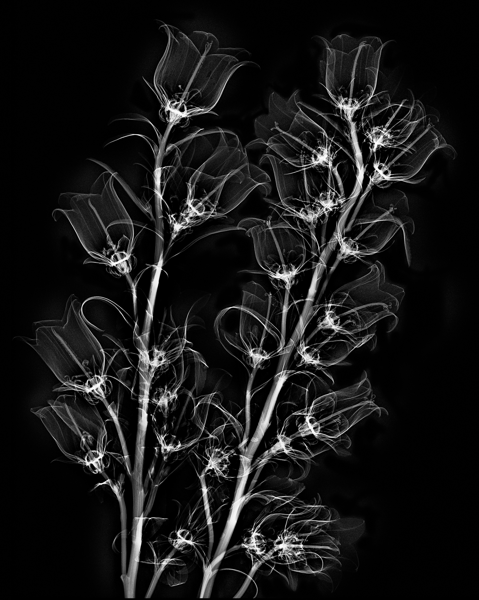
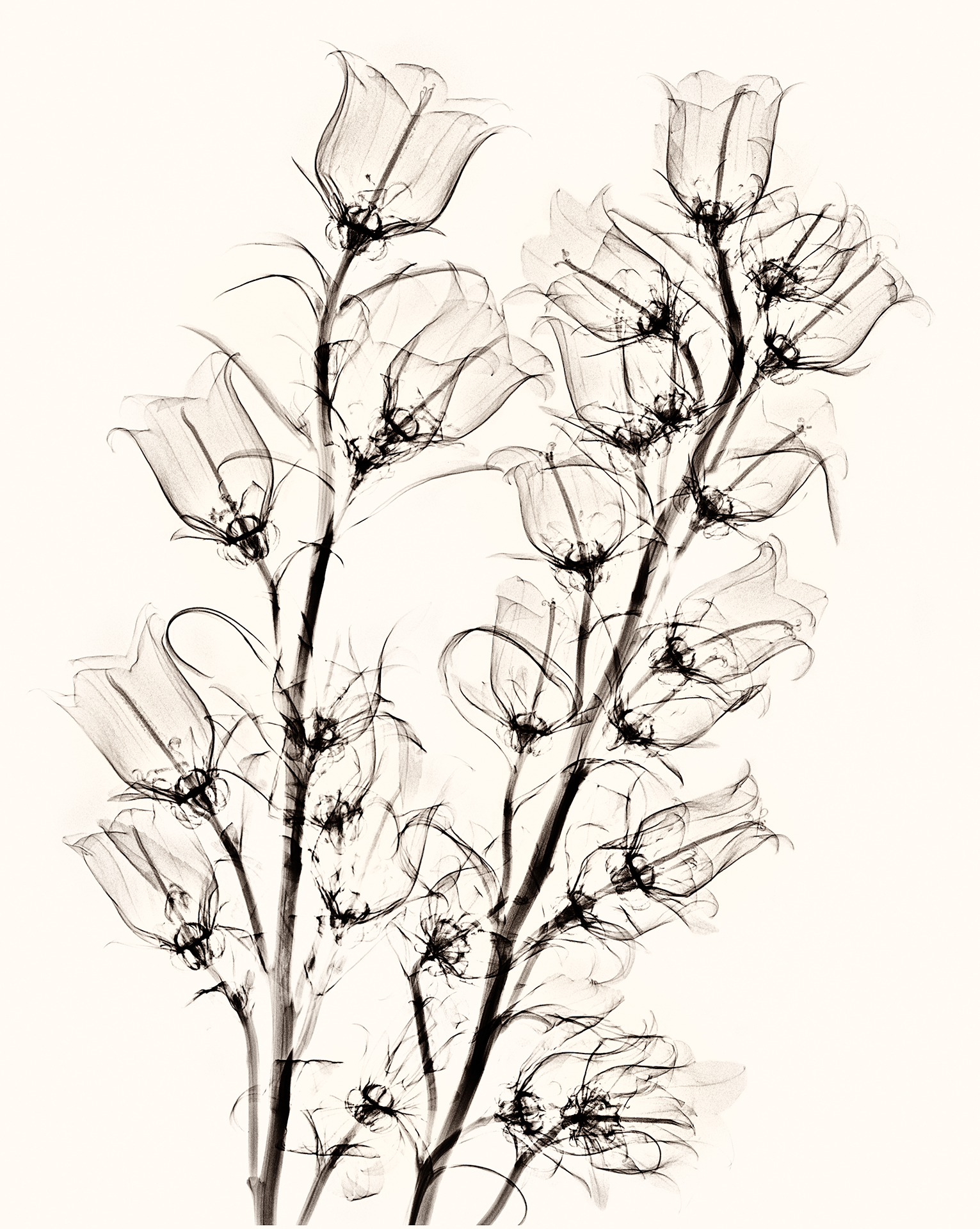
The examples so far on my blog are Campanulas X-Rays, Dahlia Fusion X-Rays and Light Box Photos, More Fusion X-Rays, and Sunflower X-Ray Fusion. Some comments on the process of making these images, what is involved technically, and where we go from here follow the image below (the on-black version of the Campanulas shown here is a good example of a fairly straight x-ray file without additional coloration or fusion with a light box image).
As you likely know, what we see are wavelengths emitted or reflected along the electromagnetic spectrum. Visible light ranges from 400nm to 750nm in terms of wavelength. In comparison, Julian tells me that the x-rays we used had a wave length of roughly 0.04nm (or shorter in wavelength by a factor of about 10,000 than visible light).
Looked at from one viewpoint, X-radiation is just another form of photography using a digital sensor to capture the data generated by exposure to the radiation from an electromagnetic wave. As with any kind of photography (or, indeed, any human endeavor), one of the key ways to get better is to practice a great deal (after all, practicing is how musicians get to Carnegie Hall).
Julian and I had three sessions of x-ray photography of flowers, and by the third session we were beginning to get the hang of the thing.
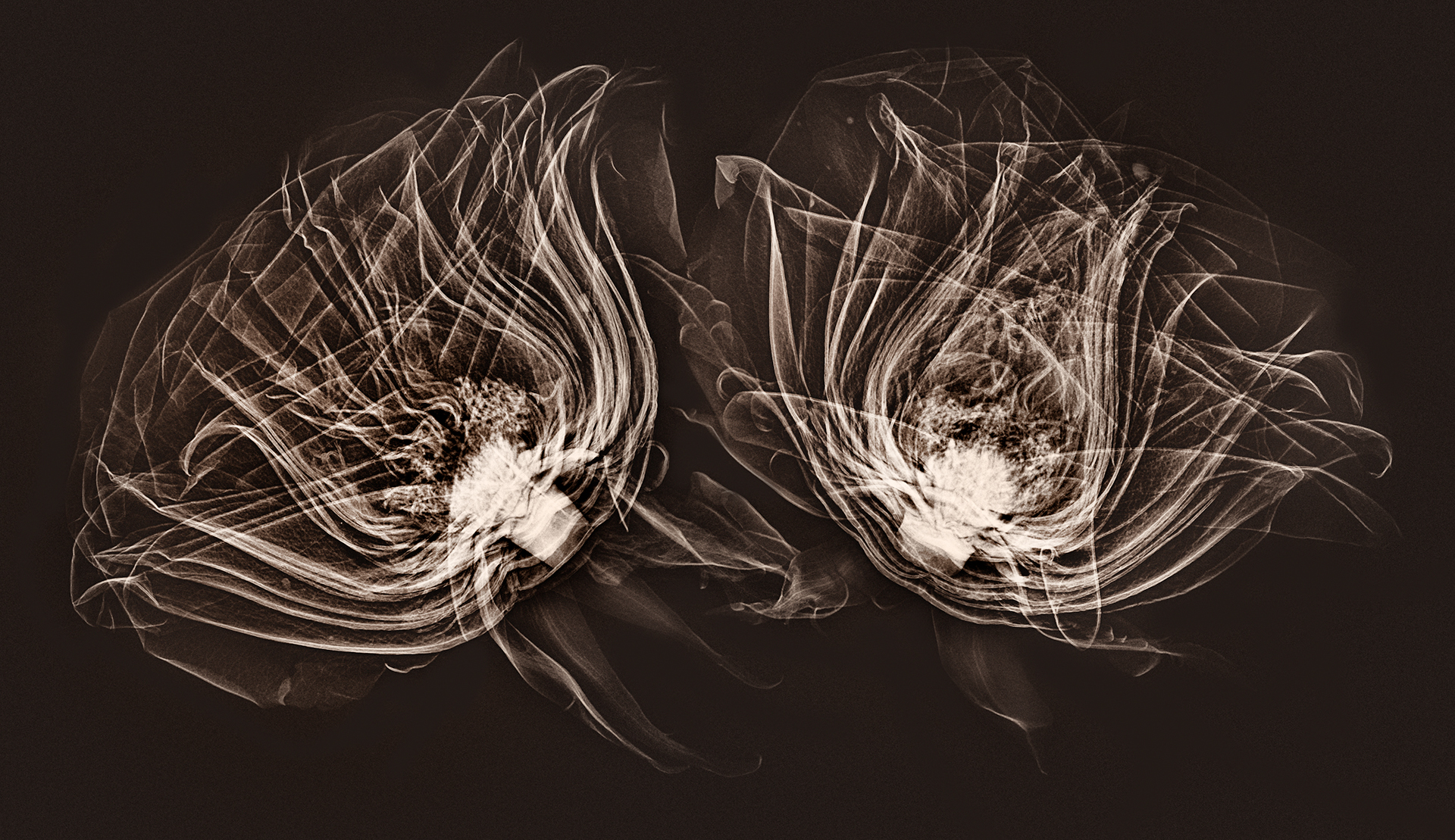
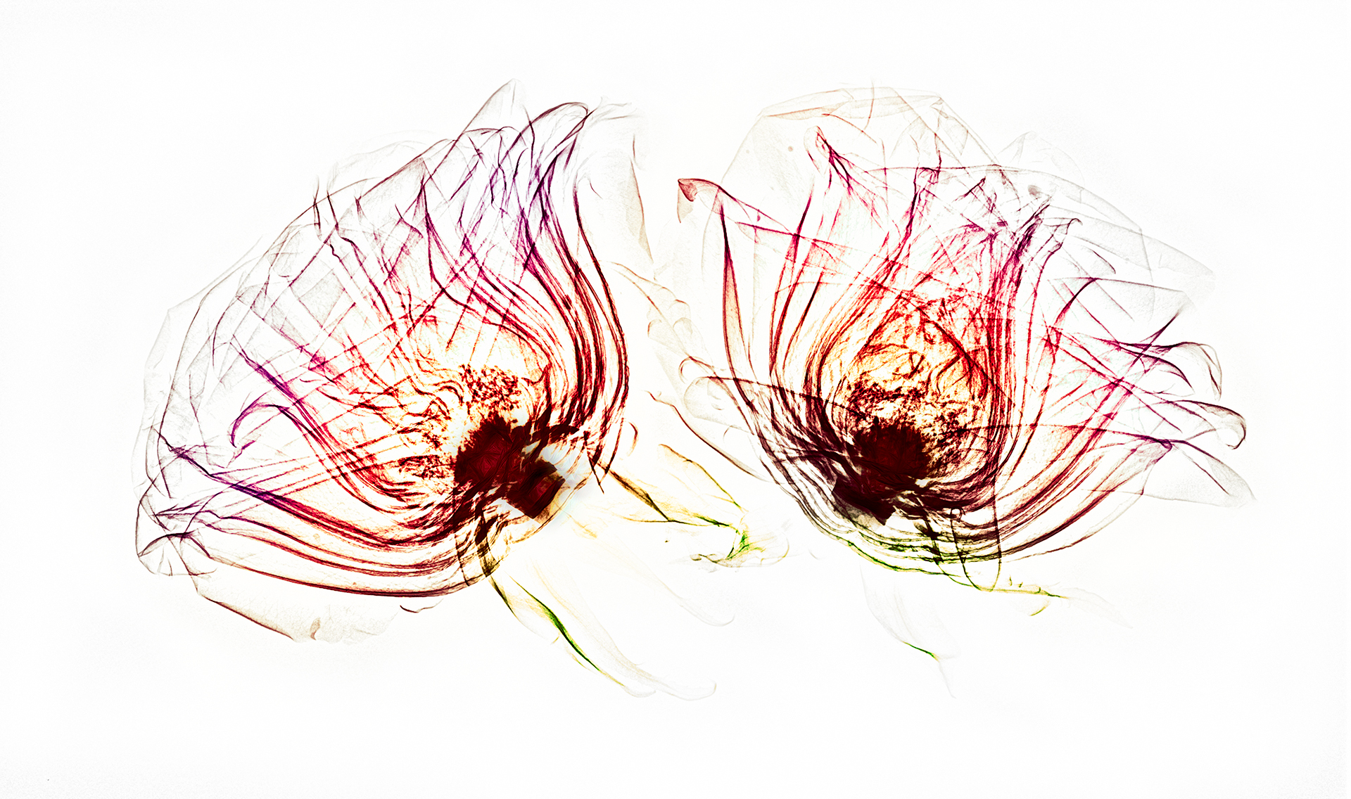
Our initial idea was to blend x-ray captures of flowers with light box flowers for transparency images. This “fusion” would allow us to show the inside and the outside of the flower composition. We used a clear and rigid plexiglass sheet to align the flowers for both processes.
The medical x-ray process involves both generating the x-radiation, and capturing it on a digital sensor. In this sense, it is analogous to firing a studio strobe and capturing the light waves emitted on a standard camera sensor. With the stationary medical x-ray device, I was reminded most of an old-fashioned analog darkroom enlarger, where the light beam from the enlarger is captured on media directly below it (photographic paper that is sensitive to light in the analog darkroom process).
We used an x-ray machine designed for mammography for these images.
What turns out is that x-radiation has very different properties as the electromagnetic source than the visible spectrum. X-rays both scatter and decay, in a way that the visible light we are used to does not. So to get good x-ray compositions, it was necessary to work with the characteristics of how the x-radiation emitted from the mammogram system would be captured by the digital sensor, and to arrange the flower compositions accordingly.
To put this comprehensibly, the mammogram system was designed to best capture the shape of the human female breast. We got better results to the extent that our composition could mimic this three-dimensional shape and positioning.
After arranging the flowers on the plexiglass, and aligning the plexiglass with marks we had set up on the machine, Julian and I would huddle behind the operator’s leaded glass shield as Julian operated the machine.
The straight x-ray photos of flowers are really beautiful, and I am grateful to have had the chance to experiment with this. I am also very excited about the fusion imagery, which I have never seen before. I look forward to exploring this area further, and to more collaboration with Julian.
Peter
31 Aug 2018Looks like lots of fun. I would be interested to see photos of you two actually taking the x-ray image and the visual light images. Also how do you get the visual image to exactly register with the x-ray image.
Harold Davis
1 Sep 2018More on this topic later. Yes, we do have some photos of working with the x-ray equipment. In terms of alignment, we placed the flower compositions on a sheet of plexiglass, with the x-ray machine borders marked off. We then moving the plexi (with the flowers) from x-ray system to light box (or vice versa). Even so, the alignment was not perfect, and requires some Edit > Transform work in post-production.
Linda Perdue
24 Sep 2018A very different perspective on taking photographs of flowers! I’d love to do something like this, but I don’t have access to an x-ray machine 🙂 I agree with Peter, it would be interesting to see you doing this process – getting a better understanding of the entire process.
Harold Davis
24 Sep 2018Some more info about this in my FAQ, http://www.digitalfieldguide.com/faqs/x-ray-photos-of-flowers, and also here’s a gallery of the x-ray images.
Pingback: Postcards from Berlin